Imaging capability
Ultra-high field for more information.
5T MRI, specific protocols and imaging engineering to make the most of resolution, contrast and tissue characterization capabilities.
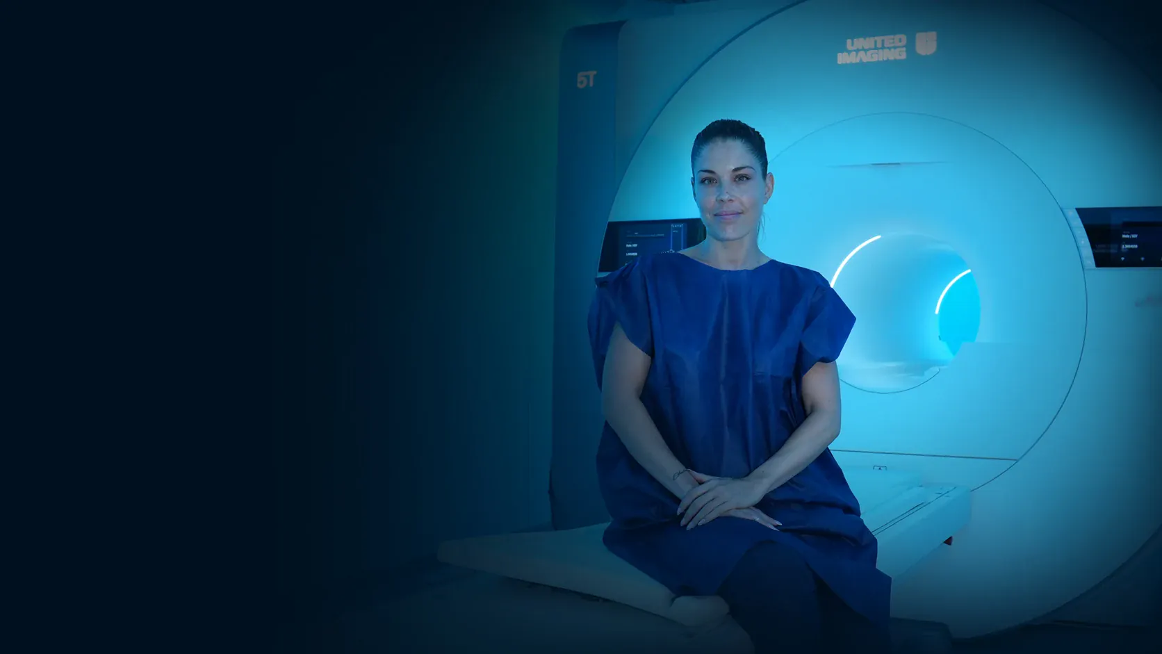
5T MRI UNIT · ULTRA-HIGH FIELD · MADRID
Never seen before.
Ultra-high-field MRI, where technology, medical knowledge and clinical experience come together to push the limits of diagnostic imaging.
HT Medica's 5T MRI Unit expands the possibilities to observe in greater detail, study with more information and better understand the human body and disease.
Más capacidad de imagen. Más conocimiento para el especialista. Nuevas posibilidades para el diagnóstico y para los pacientes.
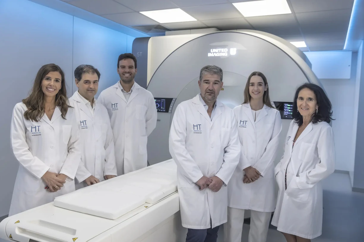
The specialization that gives imaging its value
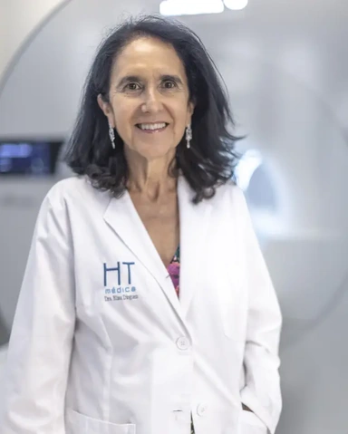
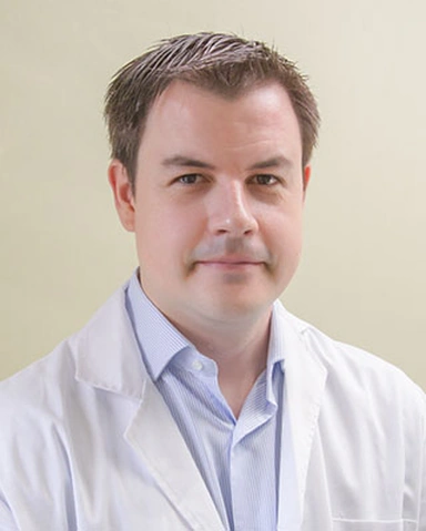
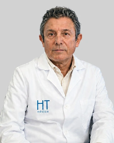

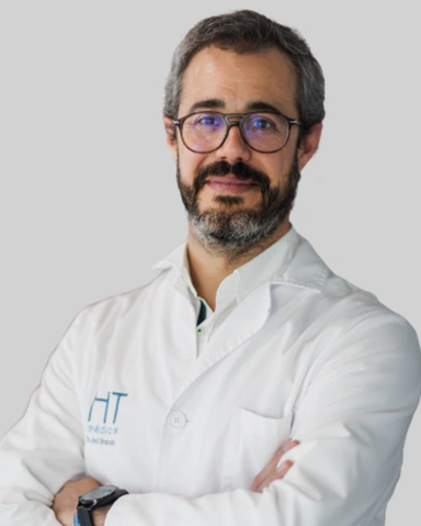
More information only has value when we know what to look for, how to interpret it and how to turn it into useful knowledge for the diagnosis and for the patient.
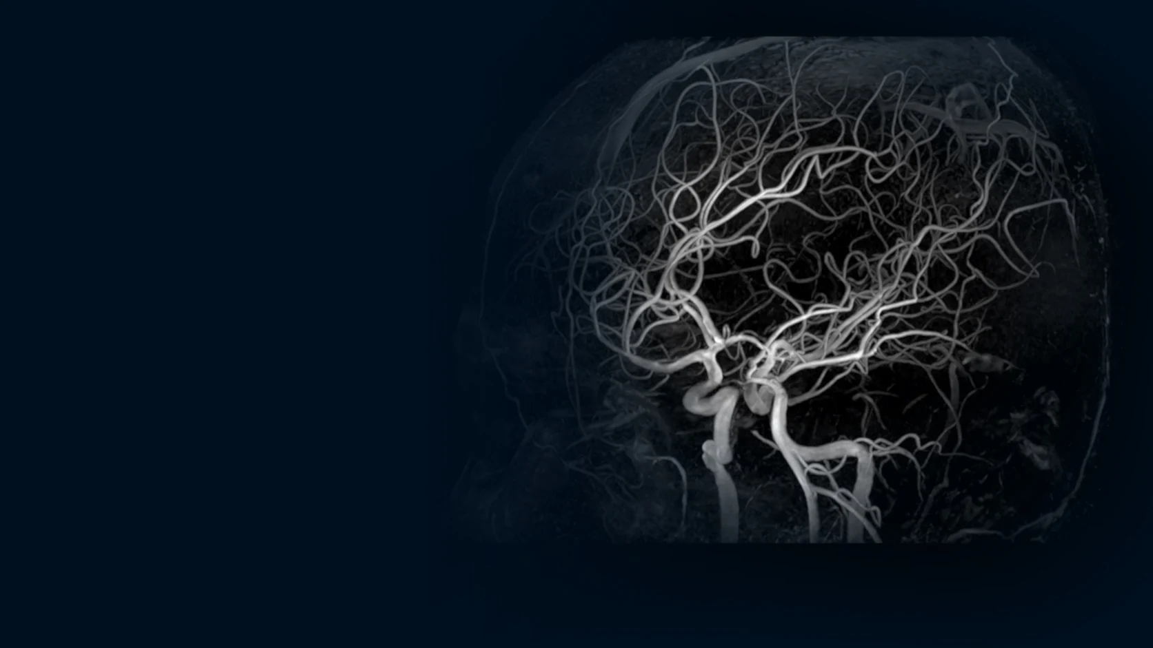
5T MRI Unit - HT Médica
Ultra-high field expands the available information. Its true value emerges when that capacity is integrated with medical knowledge, subspecialization, specific protocols, imaging engineering, artificial intelligence and research.
HT Medica's 5T MRI Unit brings all of these elements together to turn greater technological capacity into clinically relevant information for the diagnosis and for the patient.
Imaging capability
5T MRI, specific protocols and imaging engineering to make the most of resolution, contrast and tissue characterization capabilities.
Clinical capability
Subspecialization, clinical experience, artificial intelligence, research and evidence to interpret each finding in its context and answer a specific clinical question.
Subspecialized radiologists and highly qualified professionals to interpret imaging from a deep knowledge of each organ and system.
5T MRI and advanced resolution, contrast and tissue characterization capabilities.
Sequences, acquisition, reconstruction and post-processing tailored to each area and clinical question.
Advanced tools to develop, optimize and expand the Unit's capabilities.
A clinical and scientific platform to generate evidence, validate new applications and expand knowledge about 5T MRI.
More information. More knowledge. Better decisions.
5T MRI expands the possibilities of diagnostic imaging across multiple clinical areas, providing greater anatomical detail, characterization capacity and new information to answer specific clinical questions.
Clinical areas
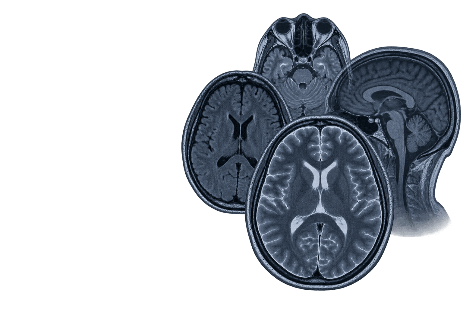
Ultra-high field makes it possible to study the brain with a level of detail that is especially relevant in complex pathologies and clinical situations. Protocol selection and specialized interpretation are essential to turn that greater imaging capacity into clinically useful information.
Clinical areas
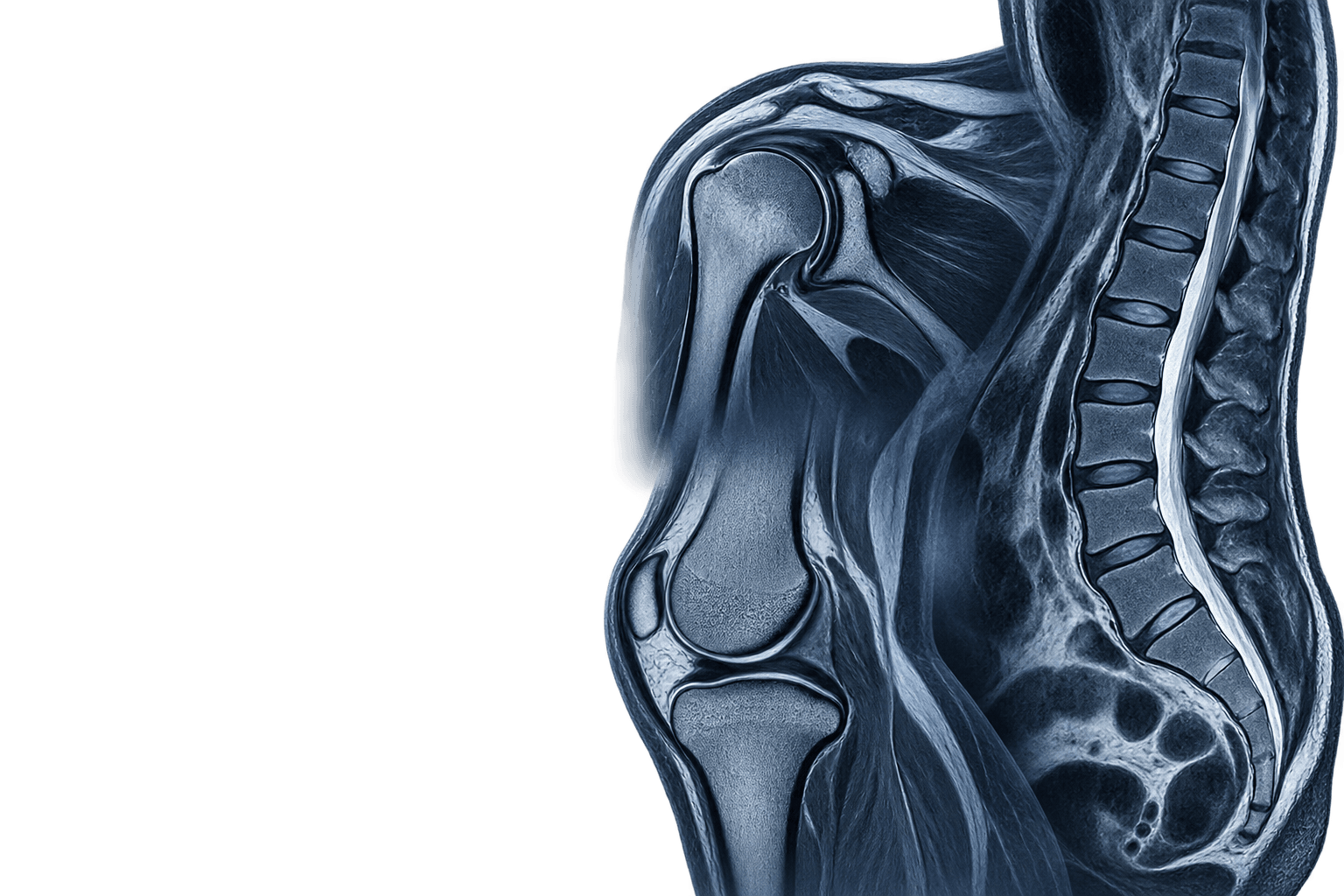
Ultra-high field allows joints, spine and muscle-tendon structures to be studied with protocols tuned to the clinical question and the context of each case. The assessment is integrated with the clinical history, the examination and the available tests to guide specialist reading.
Clinical areas
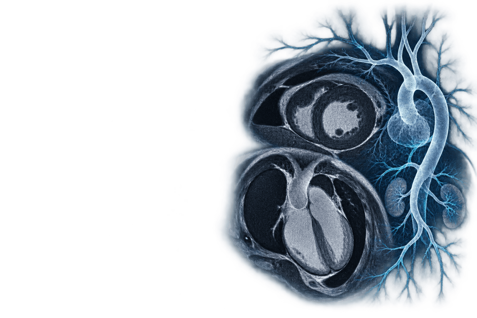
Ultra-high field makes it possible to address the heart, the great vessels and the vasculature with sequence planning geared to the clinical indication. Study preparation is adapted to the diagnostic goal, the acquisition pace and the prior information available.
Clinical areas
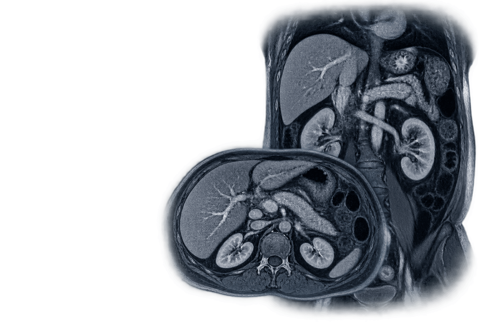
Ultra-high field allows abdominal organs and characterisation questions to be studied with protocols adapted to the organ, the system and the clinical context. The imaging strategy is defined by the indication and interpreted within each person's care pathway.
Clinical areas
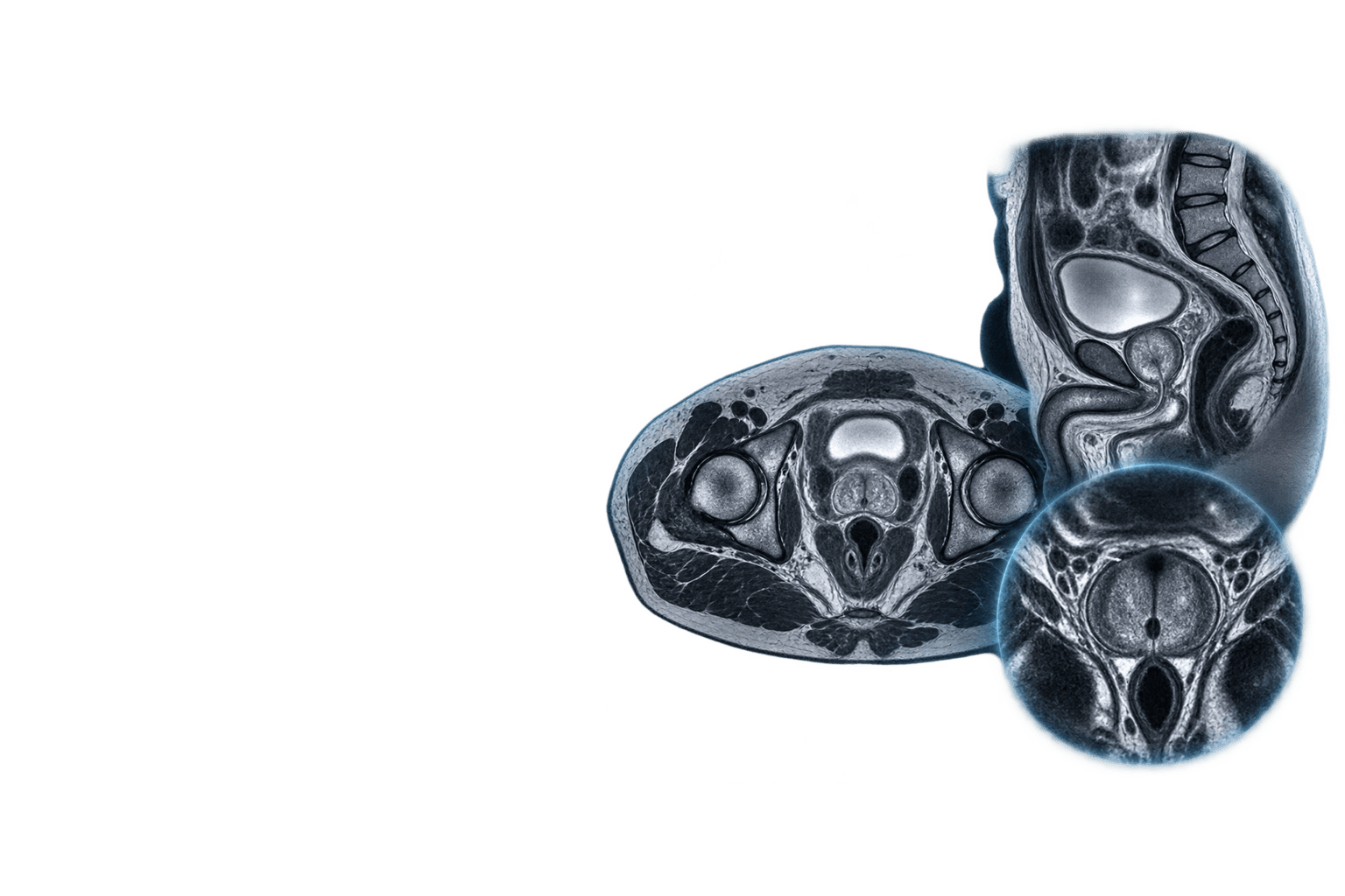
Ultra-high field allows pelvis and prostate to be studied with protocols defined by the indication, the anatomy of interest and specialist reading by organs and systems. Sequence selection is integrated with history, previous scans and the stated clinical goal.
Clinical areas
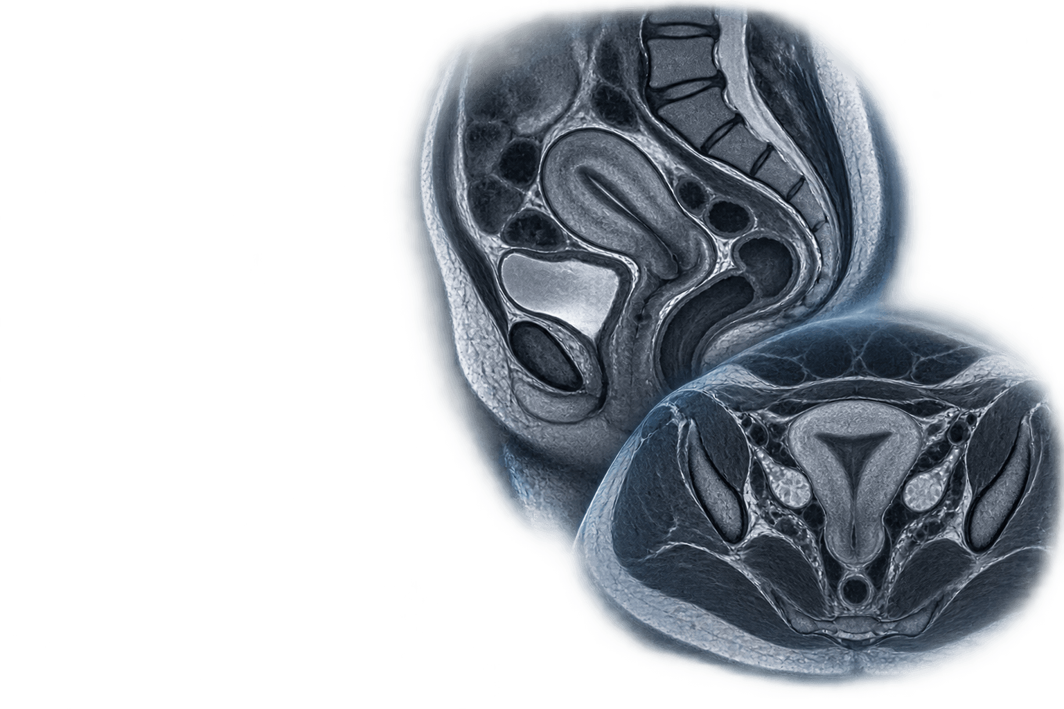
Ultra-high field allows the gynaecological anatomy to be studied with protocols adapted to the clinical question and the specialist assessment of each case. Imaging information is contextualised with the history, indication and appropriate follow-up.
Clinical areas
Ultra-high field can provide anatomical and characterisation context in selected protocols, within oncological assessment and clinical follow-up. The usefulness of each scan is assessed individually with the indication, the history and the team responsible for the case.
The potential of 5T MRI, applied to each clinical question. Specialization by organs and systems makes it possible to interpret imaging in context and turn greater information capacity into useful knowledge for the diagnosis.
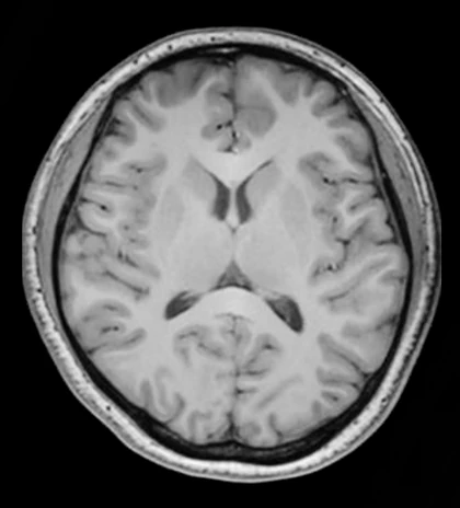
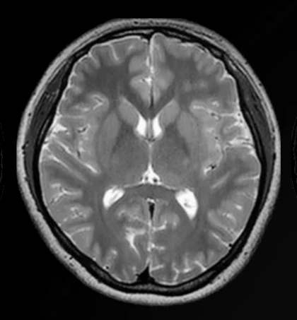
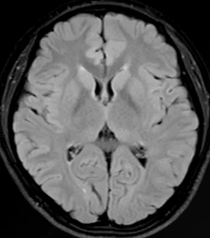
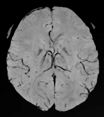
3T vs 5T · Neuroimaging
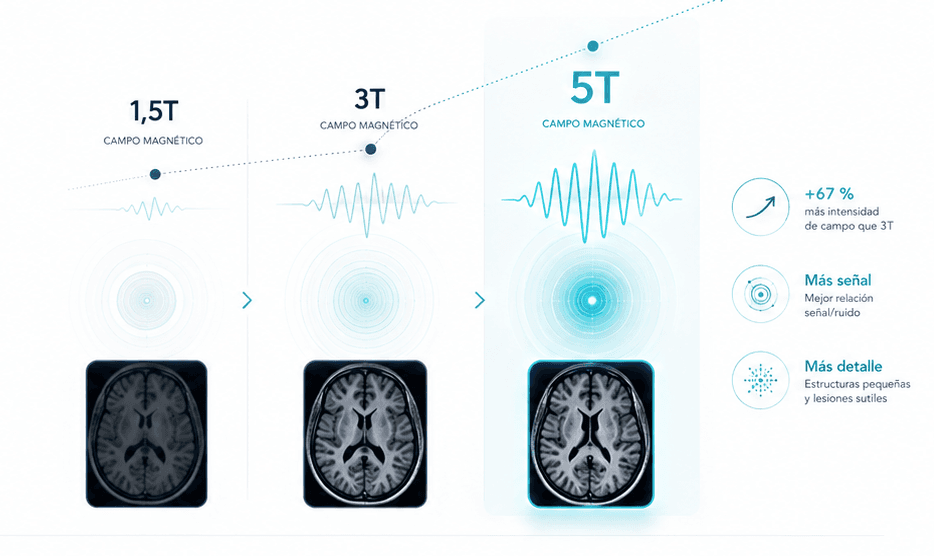
The greater signal available in 5T MRI makes it possible to work with higher resolution and anatomical definition, expanding the capacity to visualize small structures and subtle findings.
3T3T · Magnetic resonance
5T5T · Ultra-high field
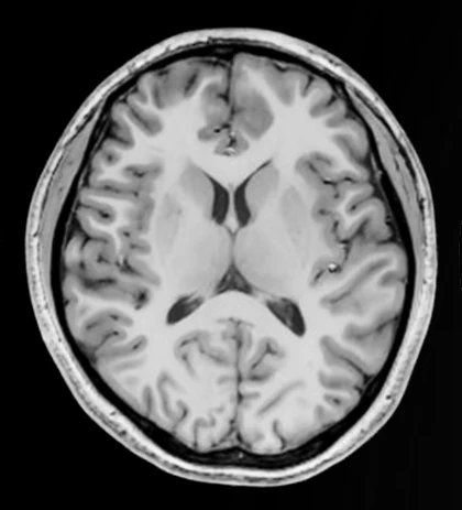
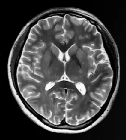
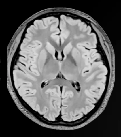
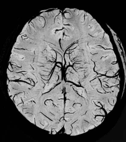
Greater capacity to visualize small structures and go deeper into the study of brain anatomy.
Greater capacity to differentiate between tissues and structures, useful to go deeper into the characterization of subtle findings.
A greater imaging capacity that, integrated with the specialist's experience, can provide additional information for clinical assessment.
More imaging capability opens new possibilities for diagnosis.
Patient experience
HT Medica's 5T MRI Unit has been designed to also care for the patient experience. Technology, privacy, comfort and efficiency come together in an environment designed to support every examination with greater calm and attention.
A carefully designed environment to offer more personal attention and preserve the patient's privacy.
Renovated spaces designed to foster a calmer experience during the examination.
Coordinated processes to ease the patient's journey and offer more agile care.
More science for fuller lives
Information and guidance for patients and families.
INFORMATION FOR HEALTHCARE PROFESSIONALS
5T MRI can provide additional information in selected clinical situations, especially when a previous study has not been conclusive, a finding requires further characterization or anatomical detail may be relevant for decision-making.
RM 5TMore detail for a future of precision
and a clinical suspicion persists that requires further assessment.
to provide additional information that helps guide the next clinical decision.
for diagnosis, treatment planning or follow-up.
in selected situations where a greater imaging capacity can provide additional clinical value.
The 5T MRI Unit team can help you assess the indication and define the most appropriate protocol for each case.
More knowledge for better decisions
Access and location
Indication and referral
To guide an enquiry, provide the clinical question, previous scans and reports, and any relevant history.
Innovation and evidence
The Unit's public documentation lets you follow its technical preparation, scientific participation and the external evidence that contextualises the 5T MRI's development.
Neuroimaging gallery
Structure ready to incorporate an authorised real case with three images and a reviewed clinical reading. In the meantime, published reference figures are shown.
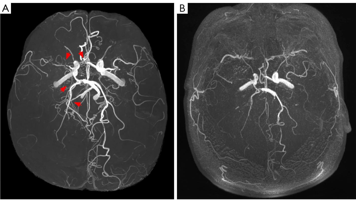
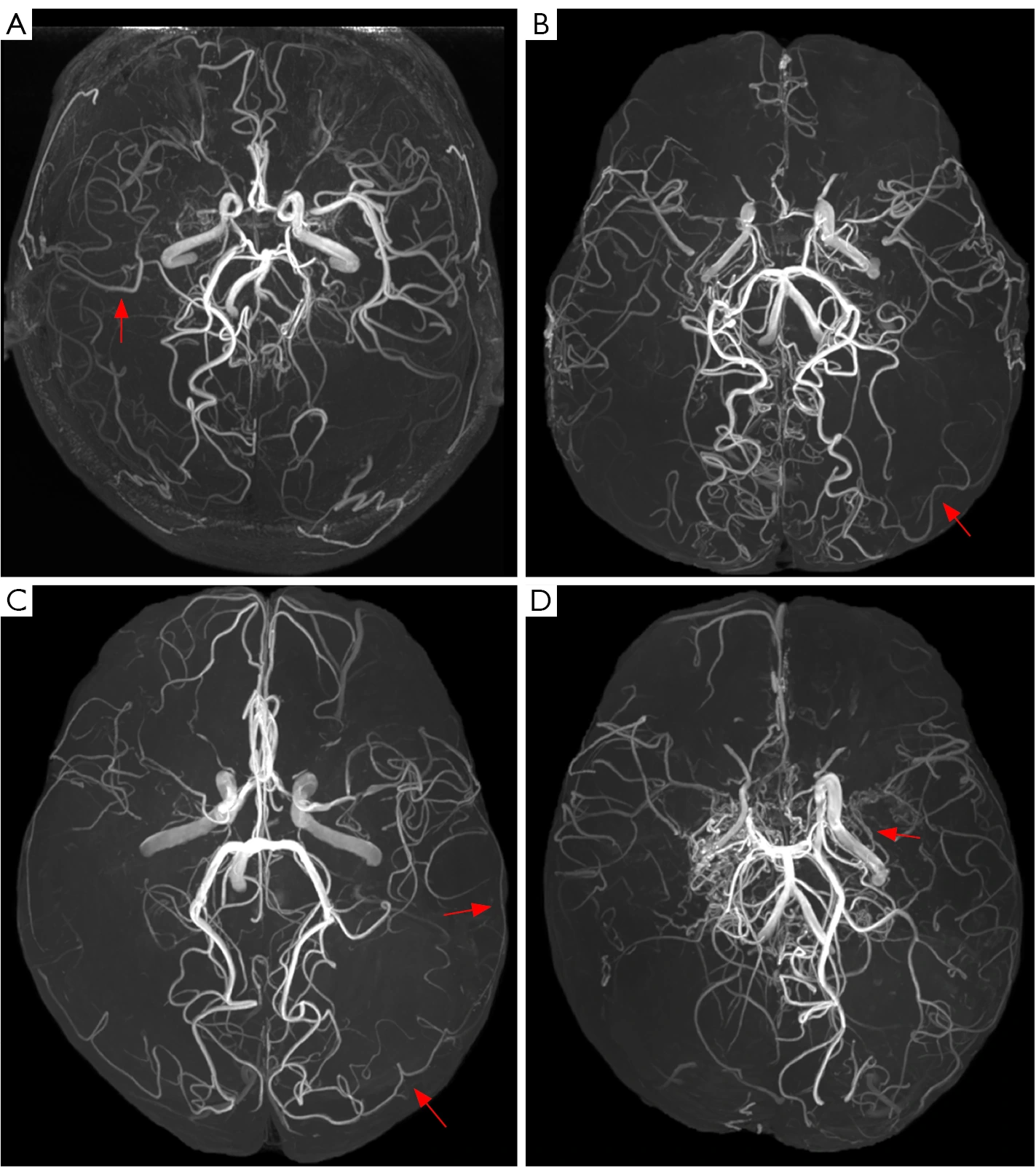
Collateral circulation comparison
Published figure of a person with headache and moyamoya angiopathy: the 5T and 3T views are shown in the same panel.
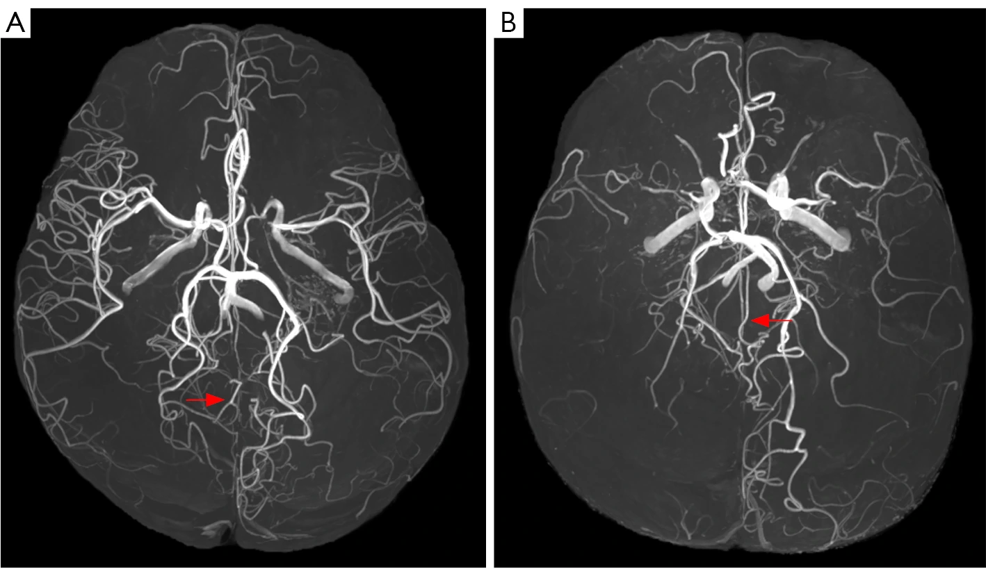
Posterior leptomeningeal circulation
Supporting figure from the same study: examples of anastomoses between PCA and ACA territories described by the authors.
Figure 1 · Published 5T/3T comparison; not an HT Médica case. · Zhou et al., 2025 · CC BY-NC-ND 4.0.
Figure 2 · Published figure; not an HT Médica case. · Zhou et al., 2025 · CC BY-NC-ND 4.0.
Figure 3 · Image from the published cohort; not an HT Médica case. · Zhou et al., 2025 · CC BY-NC-ND 4.0.
HT Médica's experience in ultra-high field is shared at symposia, professional meetings and specialist media, contributing to the conversation on the future of diagnostic imaging.
Highlights
5 highlights; scroll the rail to browse them.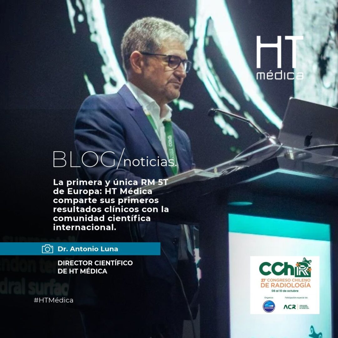
El Dr. Antonio Luna compartió la experiencia de HT Médica con la primera y única resonancia magnética de 5 Teslas de Europa, destacando los resultados preliminares en epilepsia focal refractaria y las nuevas posibilidades que ofrece esta tecnología en diferentes áreas clínicas.
View highlight →HT Médica
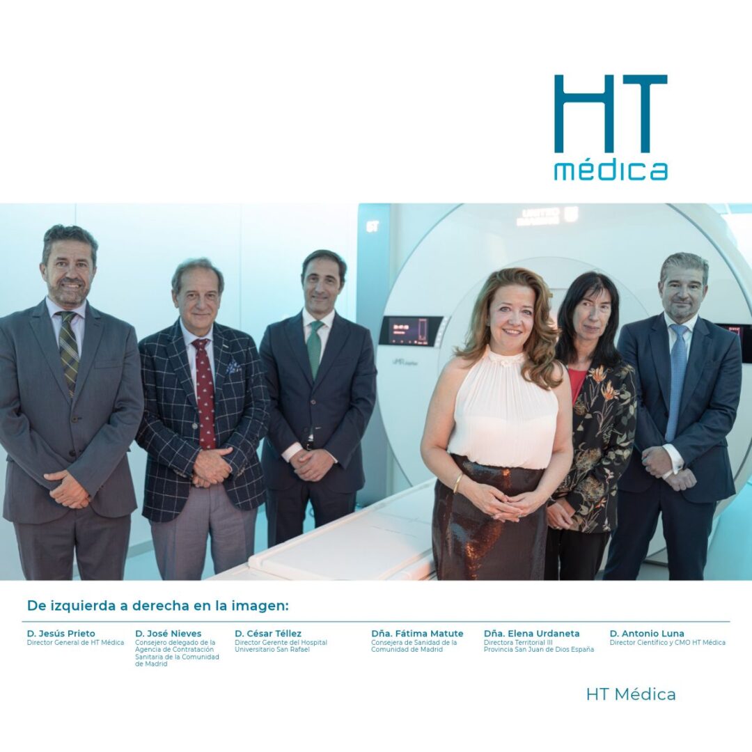
Una de las principales áreas de desarrollo será la Neurología, donde su mayor capacidad de imagen aportará un valor clínico diferencial en patologías como la epilepsia, la esclerosis múltiple o la enfermedad de Parkinson.
View highlight →HT Médica
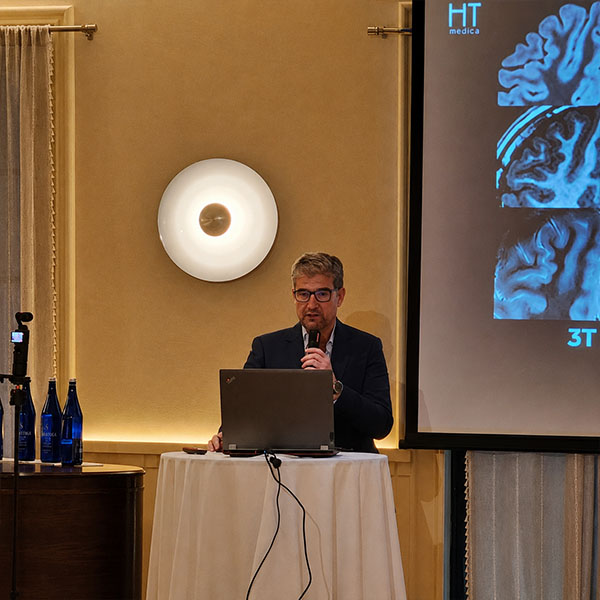
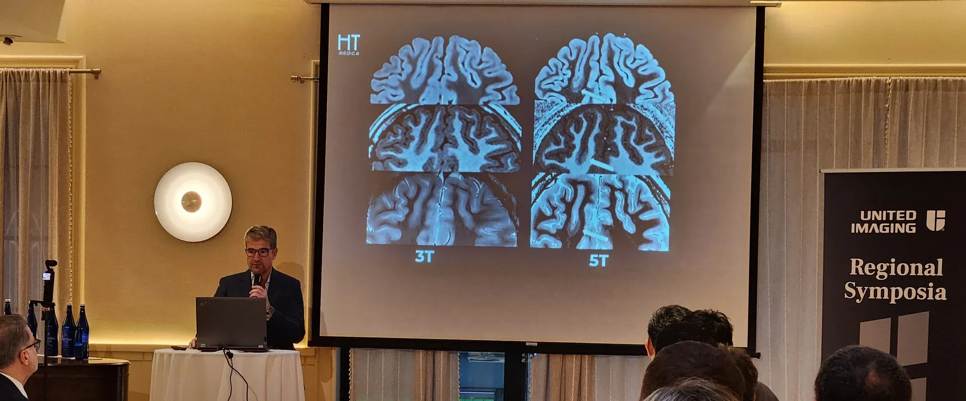
Dr Antonio Luna presented HT Médica's experience with 5 tesla MRI and its clinical potential in New York, Chicago and San Francisco.
View highlight →HT Médica
Photon-counting CT (PCCT) and 5 tesla MRI (5T MRI) represent two of the most advanced lines of technological innovation in the field of…
View highlight →HT Médica
HT Médica’s 5T MRI Unit is in its final phase of technical integration and protocol optimisation, opening a new stage for advanced medical imaging. There are advances that…
View highlight →HT Médica
News and research
The latest news from HT Médica on the Unit, 5T technology and its professionals' scientific activity.
5T Magnetic Resonance13 AGO 2026
Dr Antonio Luna presented HT Médica's experience with 5 tesla MRI and its clinical potential in New York, Chicago and San Francisco.
Read article: HT Médica's vision for the future of 5 tesla MRI at the United Imaging symposia in the United States
PCCT06 AGO 2026
5 teslas21 JUL 2026
5 teslas13 AGO 2026
5 teslas06 AGO 2026
5T Magnetic Resonance16 MAR 2026
Automatic updates · 5 Tesla RSS
Frequently asked questions
Find information about the scan first, then preparation, and finally questions about results, price and coverage.
It is an ultra-high-field MRI whose magnet operates at 5 teslas. The increased field provides more available signal and allows work with higher resolution, anatomical definition and tissue characterisation.
The 5T MRI operates at a magnetic field strength roughly 67% higher than a 3T MRI and offers additional signal capacity that can be used to optimise image quality and detail.
The greater available signal makes it possible, depending on the protocol, to work with higher resolution and anatomical definition and to study certain structures and findings in more detail.
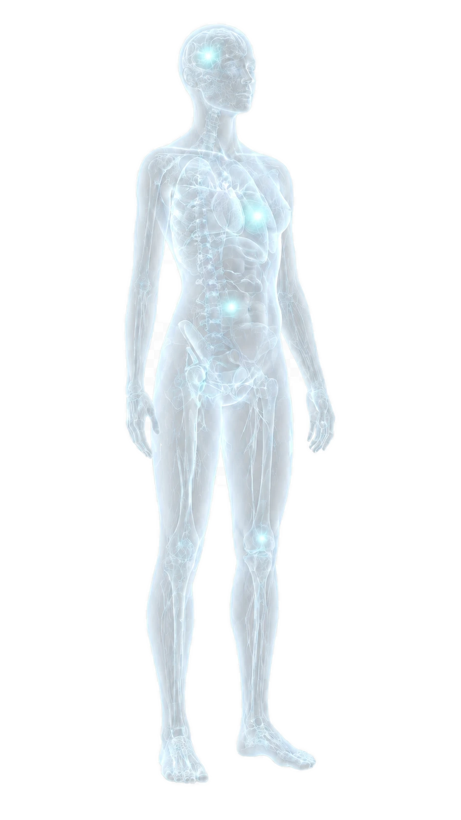
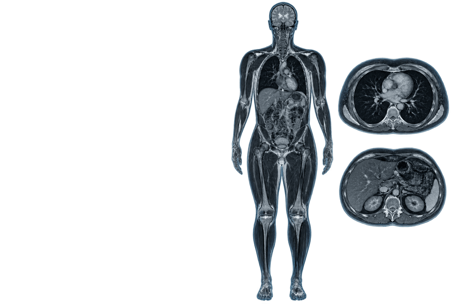
Yes. It is a whole-body platform. The Unit develops specific protocols and applications by clinical area.
In situations where a previous test was inconclusive, a finding needs further characterisation, or more detail may help answer a complex clinical question.
Yes. A 5T MRI must be indicated by a doctor.
The scan is performed in an MRI scanner. Preparation, positioning, protocol and use of contrast depend on the area to be studied and the medical indication.
Depende del tipo de estudio y de la indicación.
La compatibilidad de implantes y dispositivos debe revisarse antes de cualquier resonancia magnética.
At Hospital Universitario San Rafael, in Madrid.
Contact
Tell us what you need. Whether you are a patient, a relative or a healthcare professional, our team will route your enquiry to the right channel.
Write to uscomunicacionrm5t@htmedica.com